Review Enzymes in the degrade some types of neurotransmitters to terminate the signal.
Thủ Thuật Hướng dẫn Enzymes in the degrade some types of neurotransmitters to terminate the signal. 2022
Hà Trần Thảo Minh đang tìm kiếm từ khóa Enzymes in the degrade some types of neurotransmitters to terminate the signal. được Cập Nhật vào lúc : 2022-12-07 03:32:04 . Với phương châm chia sẻ Bí quyết Hướng dẫn trong nội dung bài viết một cách Chi Tiết Mới Nhất. Nếu sau khi tham khảo tài liệu vẫn ko hiểu thì hoàn toàn có thể lại Comments ở cuối bài để Mình lý giải và hướng dẫn lại nha.If you're seeing this message, it means we're having trouble loading external resources on our website.
Nội dung chính Show- NeurotransmittersV.A.1. Acetylcholine (ACh)Cells, Synapses, and NeurotransmittersRemoval or Destruction of the Neurotransmitter Shuts Off the Neurotransmitter SignalNeurotransmitters and Their Life Cycle☆Neurotransmitter ReceptorsNeurotransmittersWhat enzymes degrade neurotransmitters?How can a neurotransmitter signal be terminated?Which enzyme degrades the neurotransmitter norepinephrine?
If you're behind a web filter, please make sure that the domains *.kastatic.org and *.kasandbox.org are unblocked.
The influence of neurohormones and neuropeptides on the brain can be divided into two broad categories: organizational and activational.Organizational effects occur during neuronal differentiation, growth, and development and bring about permanent structural changes in the organization of the brain and therefore brain function. An example of this is the structural and organizational changes brought about in the brain by prenatal exposure to testosterone.Activational effects are those that change preestablished patterns of neuronal activity, such as an increased rate of neuronal firing caused by exposure of a neuron to substance P or vasopressin.
Numerous neuropeptides are found in the brain, where they have a wide variety of effects on neuronal function (Table 50.1). Current understanding of all the actions of neuropeptides in the nervous system is far from complete.
View chapter on ClinicalKey
Neurotransmitters
James H. Schwartz, in Encyclopedia of the Human Brain, 2002
V.A.1. Acetylcholine (ACh)
ACh is formed in a single enzymatic step. The enzyme, choline acetyltransferase, catalyzes the esterification of choline by acetyl-CoA.

The transferase is specific to cholinergic neurons and is not expressed in any other cell type. (The term cholinergic is used to denote a cell that releases ACh as a neurotransmitter. Similarly, glutaminergic, dopaminergic, and serotonergic indicate that a neuron releases glutamate, dopamine, or serotonin, respectively. If a cell responds to ACh, that cell is called cholinoceptive, a term used infrequently for the other neurotransmitters; e.g., “dopaminoceptive” is unusual.) The formation of ACh is limited by the supply of choline. Choline is not made in nervous tissue, but must be obtained through the cerebrospinal fluid from dietary sources or recaptured from the synaptic cleft from the ACh released and hydrolyzed by the enzyme acetylcholinesterase (see later discussion).
There are two general classes of acetylcholine receptors (AChR): nicotinic, responding to the alkaloid nicotine, and muscarinic, responding to the mushroom poison, muscarine. ACh is excitatory the neuromuscular junction, where it binds to postsynaptic nicotinic AChRs. As we saw with Loewi's experiment, it is an inhibitory (parasympathetic) transmitter to the heart through muscarinic AChRs. In the periphery, ACh is also the transmitter for all preganglionic neurons of the autonomic nervous system. In the brain, there are many cholinergic systems, for example, cholinergic neurons in the nucleus basalis have widespread projections to the cerebral cortex.
Nicotinic AChRs are ionotropic, meaning that, when they bind ACh, they open up to pass ions from the extracellular space into the postsynaptic neuron. Muscarinic AChRs are metabotropic. These receptors activate various second messenger pathways to produce biochemical changes within the postsynaptic neuron. Thus, as with other neurotransmitters, ACh can excite or inhibit depending on the postsynaptic receptor.
View chapterPurchase book
Read full chapter
URL: https://www.sciencedirect.com/science/article/pii/B012227210200251X
Cells, Synapses, and Neurotransmitters
Joseph Feher, in Quantitative Human Physiology (Second Edition), 2012
Removal or Destruction of the Neurotransmitter Shuts Off the Neurotransmitter Signal
Neurotransmitters bind to their receptor by mass action. This principle states that the rate of binding is proportional to the concentration of không lấy phí ligand (neurotransmitter) and không lấy phí receptor, and the rate of unbinding or desorption is proportional to the concentration of bound ligand. This is stated succinctly in the equations
[4.2.1]L+P→konL⋅PL⋅P→koffL+P
Thus, the occupancy of the receptor P with the neurotransmitter L will decrease only when the không lấy phí ligand concentration falls. Lowering the concentration of không lấy phí neurotransmitter in the synaptic gap, therefore, will shut off the continued effect on the post-synaptic cell. As shown in Figure 4.2.7, there are three general ways to achieve this end: (1) destruction of the neurotransmitter by degradative enzymes; (2) diffusion of the neurotransmitter away from the post-synaptic receptors; and (3) reuptake of the neurotransmitter either by the pre-synaptic terminal or by other cells.
View chapterPurchase book
Read full chapter
URL: https://www.sciencedirect.com/science/article/pii/B9780128008836000343
Neurotransmitters and Their Life Cycle☆
Javier Cuevas, in Reference Module in Biomedical Sciences, 2022
Abstract
Neurotransmitters are the chemical messengers that allow electrical signals from neurons to be transmitted to the postsynaptic neuron or effector target. A substance is generally considered a neurotransmitter if it is synthesized in the neuron, is found in the presynaptic terminus and released to have an effect in the postsynaptic cell, is mimicked by exogenous application to the postsynaptic cell, and has a specific mechanism for termination of its action. Various types of molecules, ranging from simple gases, such as nitric oxide (NO), to complex peptides, such as pituitary adenylate cyclase-activating peptide, satisfy these criteria. Most small-molecule neurotransmitters, such as acetylcholine and dopamine, are synthesized in the cytoplasm of the nerve terminal and transported into vesicles; a variety of substrates and biosynthetic enzymes are involved in the synthesis of small-molecule neurotransmitters. Only 12 small-molecule neurotransmitters have been identified, but over 100 neuroactive peptides have been identified. Unlike small-molecule neurotransmitters, neuropeptides are encoded by specific genes and are synthesized from protein precursors formed in the cell body toàn thân. The emerging understanding of atypical neurotransmitters such as the gases NO and CO, lipid mediators, and the phenomena of gliotransmitter action and exosomal transmission is constantly revising the understanding of what constitutes a “neurotransmitter.”
View chapterPurchase book
Read full chapter
URL: https://www.sciencedirect.com/science/article/pii/B9780128012383113182
Neurotransmitter Receptors
Richard Knapp, ... Henry I. Yamamura, in Encyclopedia of the Neurological Sciences, 2003
Neurotransmitters
Neurotransmitters are chemical compounds released by neurons after depolarization that act on other neurons to produce a response (Fig. 3). The response produced by a neurotransmitter is mediated by a neurotransmitter receptor capable of recognizing it. Neurotransmitters are the principal means by which neurons transfer information to each other. Characteristics of a neurotransmitter include its synthesis in the neuron, concentration in membrane-enclosed vesicles presynaptic terminals, release by neuron terminal depolarization, induced activity the postsynaptic terminal as a consequence of receptor binding, and removal from the synapse to terminate this effect. The defining characteristics of neurotransmitters have become less stringent due to evidence of some neurotransmitter release nonsynaptic sites and because of the properties of unusual neurotransmitter-like molecules such as nitric oxide.

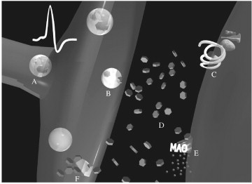
Figure 3. The synthesis, storage, action, and termination of norepinephrine, a representative brain neurotransmitter. (A) Norepinephrine is synthesized in the nerve cell and packaged into vesicles. In preparation for release, these vesicles are transported to the nerve terminal. (B) Upon arrival of an action potential the axon terminal and the resultant calcium entry, vesicles fuse with the nerve terminal membrane, thereby releasing their contents into the synapse. (C) Released neurotransmitter diffuses across the synaptic cleft and can interact with postsynaptic receptor targets to cause excitatory or inhibitory postsynaptic potentials and/or stimulate second messenger systems. Termination of the response is accomplished by removing không lấy phí neurotransmitter from the synapse. (D) Simple diffusion can carry the neurotransmitter out of the synapse, or (E) enzymes [e.g., monoamineoxidase (MAO)] can degrade or chemically modify the neurotransmitter, rendering it incapable of further action. (F) Finally, reuptake of neurotransmitter back into the presynaptic neuron or into surrounding cells can terminate the signal as well as recycle some of the neurotransmitter. (See color plate 39.)
There are many different neurotransmitter molecules (Fig. 4). They can be categorized as small molecules and much larger neuropeptides. The smallest neurotransmitter may be nitric oxide, with a molecular weight of 30, whereas the neurotransmitter peptide endorphin is composed of 30 amino acids and has a molecular weight of more than 3000—a 100-fold difference in size. Most neurotransmitters are localized to discrete parts of the nervous system, but three (adenosine, glutamate, and glycine) are present in every cell of an organism. Some neurotransmitters, including acetylcholine, norepinephrine, serotonin, and dopamine, can produce excitatory or inhibitory effects depending on the receptors on which they act. The diversity of structural and functional properties makes it difficult to categorize neurotransmitters.

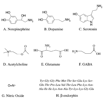
Figure 4. Examples of neurotransmitters representing the major families. (A) Norepinephrine, (B) dopamine, (C) serotonin, (D) acetylcholine, (E) glutamic acid, and (F) γ-aminobutyric acid (GABA) are small molecule neurotransmitters, where glutamic acid is also an amino acid neurotransmitter. (G) Nitric oxide is an unusual neurotransmitter in that it is an unstable soluble gas. (H) β-Endorphin is a much larger peptide neurotransmitter.
The functional properties of a neurotransmitter differ in several important ways beyond the response produced the postsynaptic site. Differences include their site of production within the neuron, the kinetics or time course of their response, and the method of removal from the synapse after release.
Small molecule transmitters, such as acetylcholine, epinephrine, norepinephrine, serotonin, and dopamine, are produced the presynaptic terminal by local enzymes. All these except acetylcholine are produced from amino acid precursors, such as tyrosine (epinephrine, norepinephrine, and dopamine) or tryptophan (serotonin). Acetylcholine is produced by the acetylation of choline, a common nutrient. Peptide neurotransmitters such as enkephalin, dynorphin, cholecystokinin, and substance P are produced by the cleavage of much larger protein precursors primarily in the cell body toàn thân of the neuron near its nucleus. The active neuropeptide products are packaged in secretory granules and then transported to their sites of release. One consequence of this difference between small and large neurotransmitters is that under conditions of high activity the neuropeptide supply the presynaptic terminal can be exhausted.
The response kinetics for neurotransmitters differs depending on the type of receptor on which they act. Neurotransmitters acting on ion channel receptors such as glutamate (excitatory) and GABA (inhibitory) produce very fast responses (milliseconds). Glutamate and GABA also act on another class of receptors referred to as metabotropic or G protein-coupled receptors. These responses are much slower and can last for seconds to hours. The response mediated by an ion channel receptor results from the flow of ions (sodium, potassium, chloride, or calcium) that occurs when the transmitter opens the channel. Responses mediated by G protein-coupled receptors occur more slowly because they result from the activation of an extended series of enzymes.
There are two principal mechanisms by which neurotransmitters are removed from the synaptic space. The majority of neurotransmitters, including all neuropeptides and many small neurotransmitters, either diffuse away from their site of release or are destroyed by enzymes present on cell membrane surfaces. Acetylcholine is a classic example because it is very rapidly destroyed by acetylcholine esterase, which hydrolyzes the ester bond between the acetic acid and choline components of the neurotransmitter. Neuropeptides are degraded into their constituent amino acids by protease enzymes. Some small molecule neurotransmitters (e.g., norepinephrine, dopamine, and serotonin) are recaptured by the presynaptic terminal through a process called reuptake. Reuptake provides a means of recycling the transmitter so that high levels of neurotransmission can be maintained.
What enzymes degrade neurotransmitters?
Degradation. Neurotransmitters can also be broken down. The classic example of this mechanism is the breakdown of the neurotransmitter acetylcholine into its constituent parts, acetate and choline, by the enzyme acetylcholinesterase (AChE).How can a neurotransmitter signal be terminated?
The neurotransmitter termination can occur in three ways – reuptake, enzymatic degradation in the cleft and diffusion.Which enzyme degrades the neurotransmitter norepinephrine?
Monoamine oxidase (MAO) is an enzyme involved in the degradation process for various monoamines released by neurons and glia cells, including DA, serotonin and norepinephrine (NE). Tải thêm tài liệu liên quan đến nội dung bài viết Enzymes in the degrade some types of neurotransmitters to terminate the signal. Diffusion of neurotransmitters Neurotransmitter degradation Neurotransmitter reuptake
Post a Comment